Roohealthcare.com – The Eustachian tube is made up of two distinct groups of muscles: the tensor and the levator. The tensor muscle opens the eustachian tube very quickly, while the levator creates tension, dilates the pharyngeal orifice, and maintains it open for long periods. As we get older, the function of the tensor muscle decreases, and the levator muscle begins to weaken.
Eustachian Tube An Important Part of Hearing and Balance
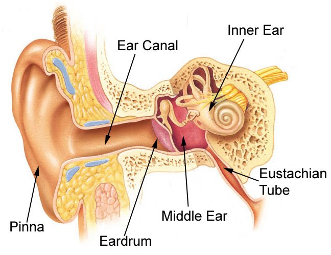
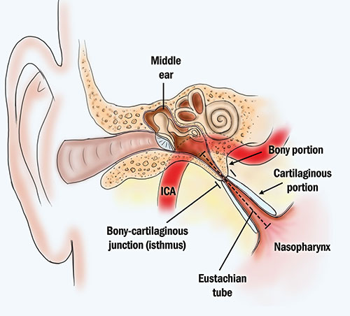
The Eustachian tube connects the middle ear with the pharynx. It is approximately 36 mm long and directs the ear downward and medially. The tube also forms a 45-degree angle with the sagittal plane. The eustachian tube contains many mucous glands and adenoid tissue. The Eustachian tube is a vital part of hearing and balance, and any obstructions can cause problems with it.
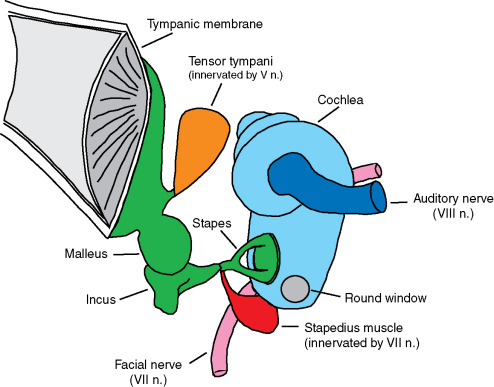
The two muscles that are attached to the Eustachian tube are called the tensor tympani and levator veli palatini. The tensor tympani originates from the superior margin of the medial cartilage of the pharynx and joins the tensor tympani and tympanic nerve. Together, these two muscles are responsible for adjusting the air pressure in the oropharynx during speech.

The symptoms associated with PET are varied. Acute symptoms may include “muffled hearing,” autophony (feeling as if someone is breathing in the ear), and a constant urge to clear the ear. In more severe cases, tinnitus or fullness or blockage of the ear may also be present. Exercise may worsen these symptoms because it increases respiratory rate and reduces blood flow to the eustachian tube.
The Eustachian Tube Protects the Middle Ear
A study of the eustachian tube revealed that it consists of two segments, a collapsible segment and a non-collapsible portion. The two pieces of muscle are connected by tubal muscles. The tensor veli palatini muscle is comprised of two functionally different groups of fibers. The fibers that open the ET are attached to the dorsal portion of the tube.
Besides being essential for airflow, the Eustachian tube protects the middle ear. During times of sudden changes in pressure, the tube allows mucus to flow out. It also equalizes pressure throughout the day. Those who have good muscle control of the tube can hear popclickity sounds or blow air through it to make their voices louder. The ear itself is a vital part of hearing health.

The TVP muscle, also known as the tensor veli palatini (TVP), originates from the base of the temporal bone. The tendon anchors into the soft palate. The muscle also operates in tandem with the tensor tympani (TT). Their innervation is similar. In addition to their joint positions, the TVP has a common tendency to lose strength as we age.
Tensor Tympani Inserts into Grip of Maleus
The tensor tympani is the larger of the two Eustachian Tube Muscles. They are attached to the malleolus in the middle and superior ear and serve to dampen sound. They originate from the cartilaginous part of the eustachian tube and the adjacent section of the greater sphenoid bone. The tensor tympani inserts onto the handle of the malleus.
The two muscles that make up the PM work together to open the tube during swallowing are connected to the mandibular branch of the trigeminal nerve. The PM is essential for peristaltic movement of the tube during swallowing and mimics the motions of the LP when swallowing is absent. In addition to opening the tube, the muscles help to maintain the shape of the soft palate. This allows them to act on the tube directly to provide greater force.

MRI is another method used to study the eustachian tube. It helps to visualize the eustachian tube and assess how it works. MRI also helps to evaluate inflammation in the middle ear in experimental animals. Further, MRI is able to evaluate the effect of botulinum toxin A on the function of the eustachian tube. So, if you think the muscles of the eustachian tube are important for healthy swallowing, you should definitely consult with a doctor.