Roohealthcare.com – If you are interested in getting a Retinal Injection, you’ve come to the right place. In this article, you’ll find information on the process and what to expect. You’ll also learn what to expect from your visit. Here are the things to consider before your procedure. Listed below are some of the most important considerations for a successful procedure. Listed below are a few common complications associated with Retinal Injection.
Procedure by Applying Pressure to Visible Subretinal Fluid
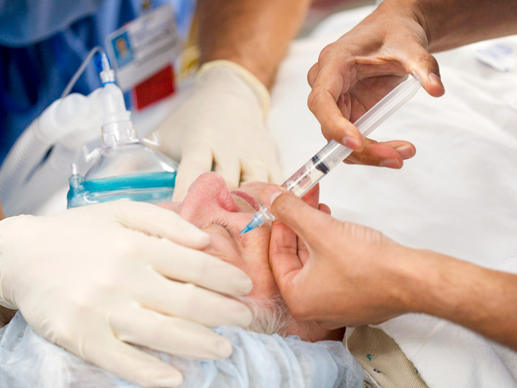
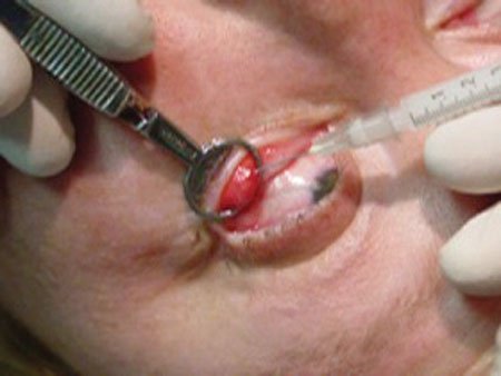
The procedure involves using a 30-gauge needle mounted on a 1-ml syringe to inject VB. The needle is then placed tangentially to the globe and inserted by applying pressure until the subretinal fluid is visible. During the procedure, your doctor will carefully guide the needle into the subretinal space. It can take up to five minutes to perform the procedure, depending on the severity of the disease.
IntactEye uses a two-view projection technique to measure the location of the injection. The center of the injection is calculated using two separate views. Currently, this method can only track one injection. It may be modified to process multiple injections of different dyes, as well as focal retrograde injections. IntactEye uses an image analysis program called Retina. It can help you see the exact location of the injection by calculating a projection of the retina onto a sphere.
In order to understand what happens after Retinal Injection, researchers first need to understand how the vector works. Injecting rAAV into the retina can enhance the efficiency of the transmission of rAAV. It will also increase the number of photoreceptors and ganglion cells in the retina. When compared to an uninjected retina, rAAV is more efficient in the retina and results in the pan-retinal spread.
Using the Injection Software System
Injections are costly. Modern retina practices have to manage the inventory of these expensive medications. An even small quantity of medication can spoil or be lost. Hence, it is critical to ensure that your staff is aware of the costs and procedures involved. Using an injection software system can reduce your costs and help you save on time. A great way to manage Retinal Injection costs is by choosing an automated system that tracks your patients’ past treatments. ArbiMed Retina helps you assign doses and select patients for a smooth procedure.
When performing Retinal Injection, the site of injection is extremely important. The site must be 3.5 to four mm posterior to the limbus. If possible, the doctor should use calipers when making the injection. The needle should be smaller than normal. Also, the needle should be tunneled into the eye before being inserted to avoid vitreous reflux. A well-positioned needle is crucial for a successful procedure.

The method for performing a Retinal Injection is straightforward and easy to learn. The more familiar you are with the procedure, the lower your chances of causing trauma to the retina. The use of a micromanipulator can reduce the traumatic impact of the procedure. OCT imaging systems are available for in-vivo monitoring and high-resolution images. They are also excellent for murine disease models. There are many advantages of using this method.
Procedures Using Two Different Approaches
In recent years, several approaches have been used to inject proteins into the retina. Adeno-associated virus (AAV) is one such virus. Adeno-associated virus (AV) is a harmless virus used in many medical treatments. The AAV particles are delivered into the subretinal space between the photoreceptors and the retinal pigment epithelium. Once the AAV-101 DNA payload has been loaded into the lower layers of the retina, it creates a “Biofactory” where proteins are synthesized.
The procedure requires two different approaches: the first involves the subretinal space, while the second involves insertion through a small slit or artificial porous substrate. A micromanipulator setup minimizes traumas and is safe for rodents. A micromanipulator allows for precise placement of the RPE cells. It can also be performed by hand. The first stage of the procedure involves preparing the rodents. A micromanipulator is used to deliver the RPE cells into the subretinal space. The other option is to use an OCT system to see the detachment of the retina.
An anti-VEGF drug injection may also be given intravitreally to patients with degenerative retinal diseases. Anti-VEGF injections can maintain or even improve the visual acuity of patients with degenerative retinal diseases. These injections require dedication from the patient and caregivers. The anti-VEGF therapy has transformed the treatment of macular edema secondary to retinal vein occlusion. However, it is not for everyone.