Roohealthcare.com – Shoulder X-rays may be used to diagnose and monitor shoulder arthritic disease. In addition to evaluating the disease’s progress, these tests can also help determine how to manage the symptoms. X-rays can reveal a variety of changes, including joint-space narrowing. If you suspect that you have shoulder arthritic disease, an X-ray of the affected area is essential.
Shoulder OA is One of the Most Common Musculoskeletal Diseases
Shoulder OA is one of the most common musculoskeletal diseases of the aging population. Radiographic findings of joint damage may indicate OA, but correlations between OA severity and pain sensation are inconsistent. This study will investigate the relationship between radiographic findings and pain sensation in patients with shoulder OA. The results of this study may be helpful for adjusting recommendations for primary total shoulder arthroplasty (PTA) procedures.
A x-ray can reveal the presence of bone spurs and a reduction in the joint space between the ball and socket. These are signs of shoulder arthritis, but treatment options may depend on the cause. In mild cases, anti-inflammatory medication and gentle shoulder stretching exercises may be sufficient to ease symptoms. But for severe cases, you may need to see a doctor. So, what’s the most important thing to look for in a Shoulder Arthritis X-ray?
 Symptoms of shoulder arthritis can affect a person’s daily life. If you are in pain every time you try to do something that you love, you should reconsider. If the activity is causing your shoulder pain, it is likely that you should modify your activity and seek medical attention. This will help you avoid unnecessary risks of joint damage and improve your quality of life. If you love playing golf and are experiencing symptoms, you may need to reduce the frequency of your golf game.
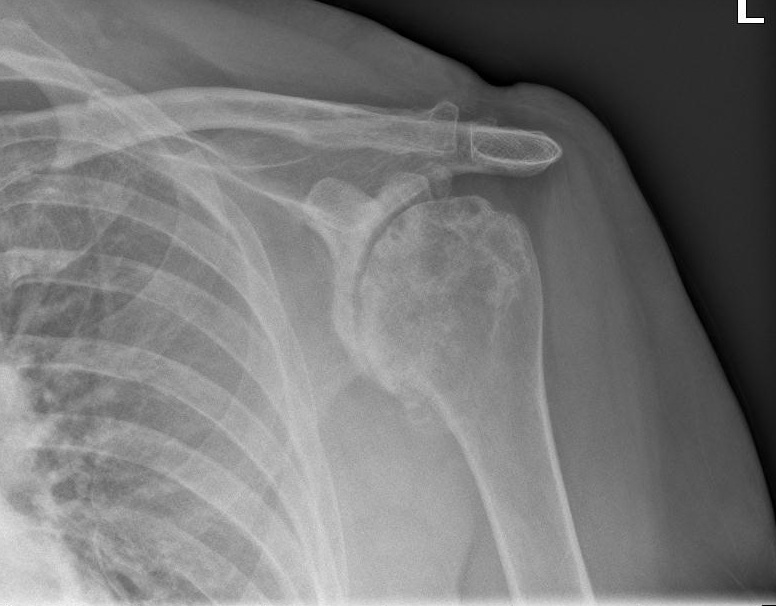
X-rays are Very Important to Identify the Cause of Pain
Shoulder Arthritis symptoms are similar to those of other conditions, which is why x-rays are so important for identifying the cause of pain. A detailed image of the joint helps doctors differentiate between osteoarthritis and other causes of shoulder pain. The space between the bones in the shoulder joint can show evidence of cartilage erosion. This means that shoulder arthritic pain is caused by a degeneration of the cartilage, and an x-ray can help them find a diagnosis.
In addition to X-rays, a doctor can also perform arthroscopy to identify loose bodies and remove inflamed joint lining. During arthroscopy, loose pieces of cartilage, bone, and tissue can be removed from the joint. A small incision is made to view the joint through an arthroscope, which is a thin camera and light source. A physician will then separate the upper arm bone from the glenoid socket. Once the shoulder joint is exposed, a metal ball with a stem is gently pressed into the humerus bone.
Shoulder Arthritis is caused by several different types of conditions. Osteoarthritis is the most common form of arthritis, and it is associated with accumulated joint damage, while rheumatoid arthritis is an inflammatory condition that results in severe damage to the joint’s cartilage. It accounts for less than ten percent of cases of shoulder arthritis and typically affects people in their early fifties.
Physical Therapists Can Help Patients Do Shoulder Exercises
MRI and CT scans are both x-rays of the joint. MRI scans are more detailed than X-rays. They can show soft tissue damage and pinpoint if a patient needs to undergo surgery or other treatment. Treatment options may include lifestyle changes, medications, or a combination of these. Shoulder exercises are a valuable part of any treatment plan. A physical therapist can help a patient perform these exercises.
Although an X-ray cannot diagnose the disease, it can reveal the presence of cartilage degeneration. In a mild form of shoulder arthritis, there may be no pain or swelling, but in a more advanced stage, the articular cartilage wears away and bones rub against each other. The resulting bone-on-bone contact can restrict movement and cause pain. Shoulder Arthritis X-rays can be an important tool in determining the cause of pain and disability.

Shoulder Arthritis X-Rays can provide valuable information on the disease’s progression and possible treatments. Fortunately, most people with the disease can manage the symptoms and remain active. The shoulder joint is composed of two bones – the glenohumeral joint and the acromioclavicular joint. The glenoid socket holds the arm bone in place while the acromioclavicular joint fits into the scapula. If you wish to send your article to roohealthcare, you can check out this page!
Reference: