Roohealthcare.com – Several types of nevi exist, including Acral nevus, Dysplastic nevus, and Giant congenital pigmented nevus. Each of these nevi can be characterized by a specific combination of features. This article will explore these common types of nevi.
Types of Nevi Found on the Skin
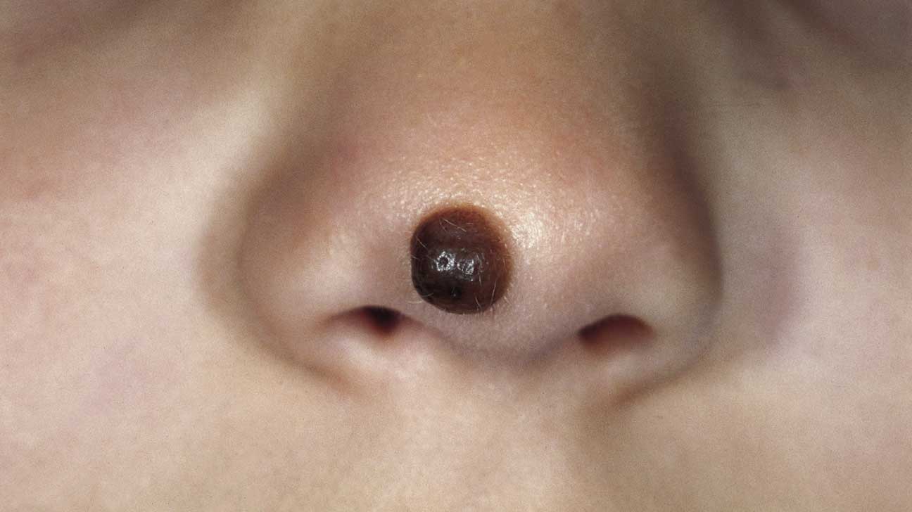
Several types of nevi are commonly found on the skin. Some of them are benign. Others are more serious. These are commonly found on the face, but can also occur on other parts of the body. They can be congenital or acquired. The risk of developing melanoma is higher for people with atypical nevi. A melanocytic nevus is a type of skin lesion that can occur at birth. The cells of a melanocytic naevus contain granular melanin.
There are three types of melanocytic nevi. They are common nevi, acquired nevi, and dysplastic nevi. Each type is classified by its architectural pattern and cellular histology. Common nevi are rounded or flat and flattened, whereas dysplastic nevi are swollen and irregular in shape. Both nevi can be light or dark, but lighter colors are more common.
Unlike a typical melanoma, a dysplastic nevus is confined to the papillary dermis. It is characterized by atypical cellular features, a predominantly nested melanocyte population, and atypical growth characteristics. These characteristics lead to a higher risk for melanoma development in individuals with dysplastic nevi.
Nevus Can Develop at Any Age
Often, dysplastic nevi appear on areas of skin that do not get much sun exposure. The nevus may develop at any age, but most atypical naevi are diagnosed between the ages of 15 and 30. It is important to note that dysplastic nevi may also develop in individuals who have had previous melanoma treatments.
Dysplastic nevi may be diagnosed by biopsy. The cells may be irregular in size and shape, with small nucleoli. They may also be spindle-shaped. In some cases, the naevus cells may mature to the base of the lesion. During the first months of life, melanoma may develop within a giant congenital pigmented nevus (GCMN). The risk of melanoma is approximately 2%-42%) in affected individuals. These lesions are typically larger than 20 cm in projected adult size.

Giant congenital pigmented nevi are usually sporadic. However, familial occurrence has been reported infrequently. In one case report, an affected infant was found to have multiple small pigmented nevi on his entire body surface. These lesions may be asymptomatic or may cause emotional distress. However, melanoma can develop during the first few years of life in affected children.
A Few Congenital Nevi Can Develop into Skin Cancer
Melanoma may also develop from melanocytes in other tissues, including the central nervous system. Melanoma may also develop in the early months of life, and a small percentage of all congenital nevi may develop into skin cancer. It is important to have periodic MRI scans to monitor these tumors and ensure proper management.
Acral nevus
Among the most important risk markers for melanoma are congenital and acquired nevi. In addition to a variety of cytologic atypia, they also may present patterns of dermal proliferation. These patterns can be confusing with atypical nevi. However, nevi at particular sites may display unique features, which may provide a clue as to their identity.

Among the most common types of nevi are the conventional nevus and the blue nevus. The conventional nevus has a densely pigmented dendritic cell component. The blue nevus is composed of a cluster of cells in the epidermis, which may have background sclerosis. These features are distinctly different from dysplastic nevi.
Reference:
Happle, Rudolf. “What is a Nevus.” Dermatology 191.1 (1995): 1-5.