Roohealthcare.com – If you’re looking for information about the anatomy of the Supraspinatus Tendon, this article is for you. It will provide the background information you need to know about this important muscle. Once you understand its role in biceps tenosynovitis, you can effectively use this muscle to perform a variety of activities. But before you learn the anatomy of the muscle, let’s first talk about its location.
The Smallest Of The Four Rotator Cuff Muscles
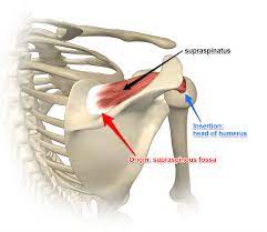
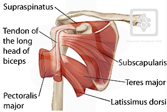
The supraspinatus is the smallest of the four rotator cuff muscles. It originates in the supraspinous fossa, a concave depression that sits above the scapula. The tendon of the muscle runs underneath the acromion and inserts on the superior facet of the greater tubercle of the humerus. The muscles of the rotator cuff work together to stabilize the glenohumeral joint, so its role is vital.
The supraspinatus muscle is made up of two components: the anterior belly and the posterior belly. If a supraspinatus muscle injury occurs, it may affect the ability to elevate the arm. During surgery, it is important to note whether the anterior belly and fibers of the supraspinatus tendon are preserved, because they will facilitate post-operative rehabilitation.
The supraspinatus tendon is surrounded by a fibrocartilage pad, which provides a transition zone between hard and soft tissue. Fibrocartilage protects the collagen fibers from sharp angulation. Moreover, this pad prevents the supraspinatus tendon from rubbing against the head of the humerus during rotation and splaying out.
Evolved to Serve a Specific Purpose on the Shoulder
The posterior belly muscle was originally more of a strap-like muscle. It was derived from the scapula’s acromion and coracoid process (central portion of the humerus). Previously, the posterior belly muscle was more a strap-like muscle and lacked an internal tendinous core. In the last few decades, this muscle has evolved to serve a specific purpose in the shoulder.
The supraspinatus tendon is prone to tearing or degenerative tears. People who engage in repetitive overhead motions such as lifting heavy objects or performing other activities that involve overhead motion should seek immediate medical attention. In some cases, supraspinatus tendon tears are asymptomatic, meaning they don’t cause any problems, but can significantly limit the range of motion of the arm.
The bicipital groove was located near the articular capsule. The anomalous structure was derived from the greater tuberosity of the humerus. It was separated from the biceps tendon by a synovial sheath. Nevertheless, the tendon structure and function were characterized by anomalies. So, if you’re looking for information about the Supraspinatus Tendon Anatomy, take a look at the following article.
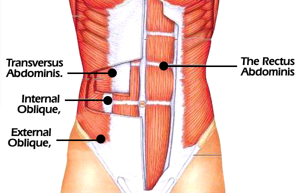
Double Layered Braided Structure and Has Two Different Abdominal Muscles
The anterior belly of the Supraspinatus tendon is thicker and asymmetrical compared to the posterior one. The anterior tendon, on the other hand, is a double-layered interwoven structure and has two distinct muscle bellies. It provides approximately 40% of the external tendon’s overall width, while the posterior tendon’s shape is thin and dispersed.
Infraspinatus muscle tears are caused by overuse or chronic instability of the shoulder. The symptoms associated with this condition include pain at night, shoulder weakness, and difficulty moving the arm during certain movements. A surgeon should be consulted if the tear is of a full thickness and requires surgical repair. If the tear is small, non-surgical treatment may be enough. However, the patient must schedule regular appointments to ensure the muscle continues to heal properly.
The Biceps tendon guides the head of the humerus. It travels along a monorail on the biceps tendon, explaining why the humerus can rotate substantially when adducted, but only slightly when maximally abducted. It is also known for its tuberosities that straddle the biceps tendon near the attachment to the supraglenoid tubercle.
Reference: