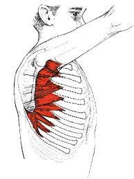
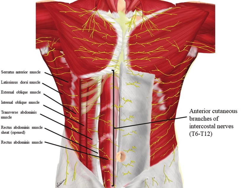
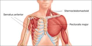
Roohealthcare.com – The Serratus Anterior Location is the area of the upper back where the scapula attaches to the thorax. Its motion is antagonistic to that of the rhomboids, which are the muscles that pull the ribs up and out of the chest. The serratus anterior has an important role in breathing, pulling the scapula forward around the thorax.
Thin Superficial Muscles in the Upper Chest Wall
The Serratus Anterior is a thin, superficial muscle on the upper chest wall. Its fibers run along the rib cage and are innervated by the long thoracic nerve, a branch of the brachial plexus. Learning how to locate your serratus anterior is easy, especially when you use mnemonics. You can try remembering the nerve roots by saying ‘C5, 6, 7 raise your arms to heaven.’ This will help you remember the correct location of the muscle.
The patient’s presenting complaint was pain and decreased strength in the left arm. She had no changes in bowel or bladder function, nor did she have night sweats. Her medications included gabapentin, omeprazole, promethazine, and ondansetron. Her symptoms were associated with a decrease in the strength of her left arm and leg. The diagnosis was serratus anterior myofascial pain syndrome and the patient consented to trigger point injections. Physical therapy was also recommended for the patient to strengthen the affected muscle.

The serratus anterior is located on the upper ribs and inserts onto the medial border of the scapula. A good reference chart can help you learn how to locate the serratus anterior. The serratus anterior performs two main functions: lateral rotation and protraction of the scapula. It also stabilizes the scapula and holds it against the chest wall.
Innervation of the Serratus Anterior Varies
The innervation of the serratus anterior varies according to its location. In the superior portion, the serratus anterior may be intimately related to the levator scapulae, and the middle and inferior segments may be true serratus anterior muscle. However, the superior portion of the serratus anterior receives innervation from two different nerves: the long thoracic nerve and the branch from C4.
Another exercise for the serratus anterior is the “wall slide.” This exercise is performed by standing facing a wall, and sliding the forearms up the wall. The shoulder blades should protract forward during the exercise. Hold the position for three seconds, then return to the starting position. Before starting any exercise routine, make sure you don’t have shoulder pain. If you have this problem, seek medical advice immediately.

In cadavers, the serratus anterior plane block is used to treat a variety of pain. In the present study, six soft-fix embalmed cadavers underwent the procedure. The serratus anterior plane block was performed by using methylene blue and latex, respectively. The latex spread was measured in three different planes: the serratus anterior, the superior-inferior plane, and the anterior-posterior plane.
Serves as a Central Control Center for the Lungs
This block is also known as a “serratus anterior plane block.” The name comes from the fact that the serratus anterior is a major nerve in the upper thoracic region. It serves as a central control center for the lungs and provides paraesthesia for the ipsilateral hemithorax. Although the serratus anterior plane block is a popular method of thoracic analgesia, it is not a suitable substitute for a paravertebral block.

The subclavius is a relatively insignificant muscle. It contributes to brachial plexus compression in thoracic outlet syndrome and is attached to the anterior scapula through fingerlike projections. The muscle has two primary functions: to protract the scapula in a punching motion and to stabilize the thoracic wall. When it is injured, however, the long thoracic nerve is compressed, causing the scapula to medial winging.
Reference: