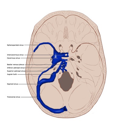
Roohealtcare.com – The importance of understanding the anatomy of the Dural Venous Sinuses cannot be stressed enough. The anatomy of this structure is crucial for draining the hemispheres. In order to perform sinus reconstruction, the anterior segment of the sinus must be preserved with a patent lumen. The recommended methods of repair depend on the mode of injury. Surgical techniques are usually used to reconstruct damaged sinuses.
Dural Venous Sinuses Diverse and Develop
The dural venous sinuses are multifaceted and develop from a widespread venous network. Understanding the anatomy of these structures is essential for interventional neurosurgical procedures and proper diagnosis. In this chapter, we review the major intramural venous sinus variants and their associated pathologies. The chapter concludes with a description of the various diagnostic and therapeutic options for patients with dural venous sinus syndrome.
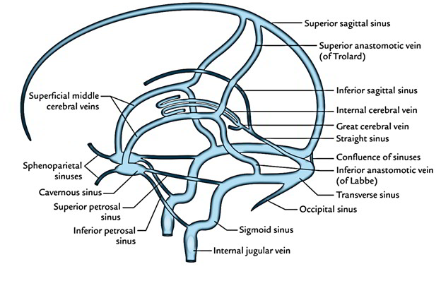
The brain contains many venous sinuses that facilitate blood flow from the brain and neck to the heart. These sinuses are divided into two groups: the superficial and the deep group. The superficial group drains the brain’s parenchyma and runs through the subarachnoid space. The deep group drains the inferior surface and carries blood to the superior sagittal sinus.

The major dural venous sinuses differ in their functions. The superior sagittal sinus (SSS) lies superior to the falx cerebri and receives blood from the cerebral veins and cerebelli veins. The inferior sagittal sinus (ISS) is the smallest of these three and lies within the inferior aspect of the falx cerebri. It connects with the great cerebral vein. The inferior sagittal sinus drains into the great cerebral vein and the transverse sinus.
The Anatomy of the DVS is More Complicated than the Arterial System
Because of the asymmetry of the dural venous sinuses, the anatomy of the DVS is often more complicated than that of the arterial system. Nonetheless, comprehensive knowledge of DVS anatomy will minimize the number of complications during treatment. The book is accompanied by illustrations and clinical photographs that illustrate the various pathological entities. The text is designed to be accessible and comprehensive and features more than 250 color images to highlight its important features.
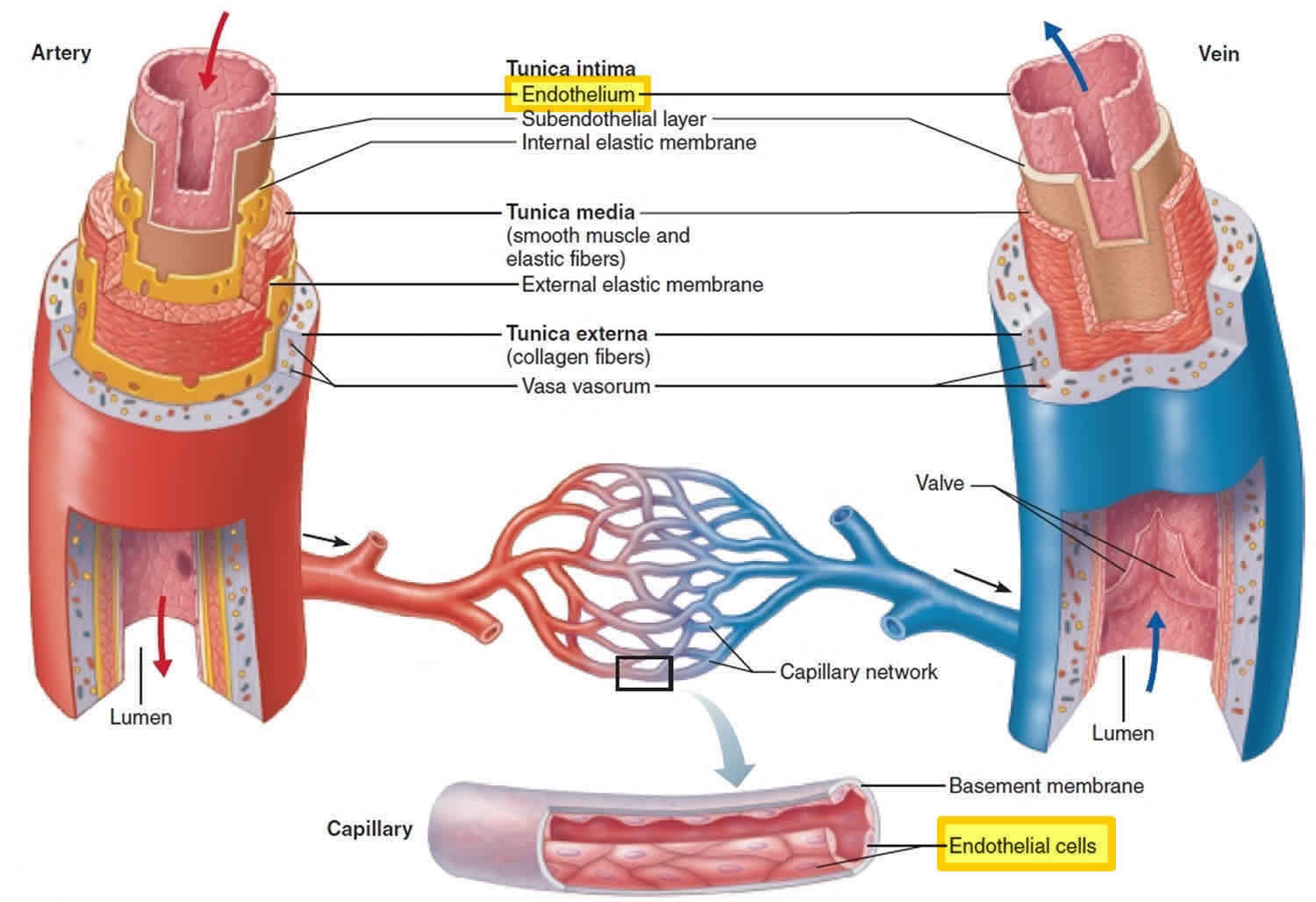
The walls of the dural sinuses are layered and highly collagenous. This structure is responsible for the high stiffness of the tissue. The walls of these sinuses are rigid and have no nerve endings, and are therefore difficult to damage. In addition to these features, there are also layers of tissue that make up the arachnoid mater membrane. These layers are the main components of the wall of the dural sinuses.

The elastic moduli of the dural venous sinus tissues range from 1.5 to 5.5 MPa. These values are considerably higher than those of other venous structures. Because of this, they should be carefully analyzed when undergoing dural sinus angioplasty. However, these figures do not fully reflect the physiology of the dural sinuses. This is why future studies should use a larger number of experimental replicates.
SSS Contains a Thin Layer of Endothelial Tissue
The SSS is responsible for the majority of cerebral venous outflow. The SSS contains a thin layer of endothelial tissue, which may not fully correspond to the anatomy of the human dural sinus. In addition, the SSS contains a largely absent falx. This tissue is therefore not a suitable animal model for the study of human dural sinus anatomy. However, pigs have been used as models for studying cerebral venous anatomy.

The superior sagittal sinus lies on the margin of the dura mater. This sinus is connected to the petrous portion of the temporal bone and the anterior clinoid process. Its margin is separated from the inferior sagittal sinus by the crista Galli fold. The inferior sagittal sinus is attached to the petrous portion of the occipital bone.