Roohealthcare.com – If you have ever wanted to learn more about the anatomy of the Pectoralis Major Muscle, you’ve come to the right place. We’ll talk about the insertion of the pectoralis major tendon and the anatomy of this muscle, and what you should look for. Here are some tips for performing an ultrasound examination of the Pectoralis Major Muscle. This is an essential muscle for bodybuilders, and it’s one of the most common abdominal muscles.
Pectoralis Major Muscle Characteristics
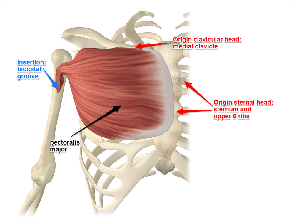
The pectoralis major muscle is a triangular-shaped muscle with origins in the sternum, clavicle, and ribs. The pectoralis major is separated from the clavicle by the external oblique fascia. It functions as both an external and internal rotator. As an additional bonus, it is a strong muscle and an excellent candidate for strengthening exercises.
A partial-thickness tear is the most common type of pectoralis major injury. The anterior layer of the pectoralis major tendon can tear under increasing mechanical loads. The remaining inferior sternal segments are also at risk of rupture. A full-thickness full-width injury involves the anterior layer of the pectoralis major tendon. This injury can even cause the clavicular head to be torn.

The pectoralis major muscle originates from the superior borders of the second through fifth ribs. Laterally, it blends with the external intercostal muscles and anterior intercostal aponeurosis. According to Anson et al., the pectoralis major muscle has coastal origins in ribs 2 through five and ribs three through four. Its origins are primarily located in the chest.
Proper Treatment of Pectoralis Major Muscle Injury
As the popularity of bench press exercises has increased, the frequency of injuries to the pectoralis major muscle has also increased. Surgical or conservative management of the injury is available, and the radiologist plays an important role in the assessment of the severity of the injury. With a high-quality MRI, the pectoralis major can be characterized with precision. However, the radiologist is an important member of the medical team and can determine the appropriate treatment.
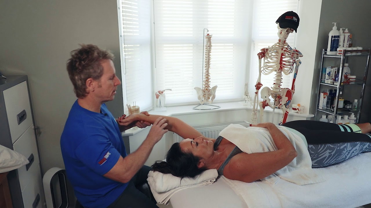
The anatomy of the pectoralis major muscle is complex and requires imaging to understand any injuries to the muscle. Regardless of the type of injury, it is important to learn about the pectoralis major muscle to ensure the best recovery possible. In addition to MRI, your doctor should perform ultrasound exams to help determine the source of the injury. These scans are useful for assessing a range of injuries to the pectoralis major muscle, so you can determine whether or not the problem is pectoral-related.

When working out, the pectoralis major muscle can be trained to do many different things. It can help your body move more quickly and effectively when you lift a heavy object. It also helps prevent injury. It is an important muscle to strengthen during the summer months. In addition to strengthening and stabilizing the pectoralis major, you can also strengthen other muscles of the body, such as the biceps and the triceps.
Pectoralis Major Receives Double Motor Innervation
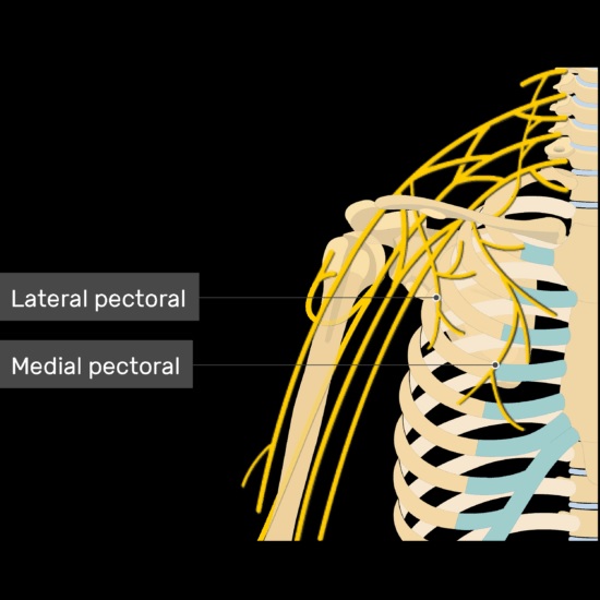
The Pectoralis major receives dual motor innervation from the lateral and medial pectoral nerves. The medial pectoral nerve innervates the sternocostal part of the muscle and extends upward into the clavicular head. The lateral pectoral nerve enters the muscle from the lateral cord of the brachial plexus. Branches of the medial pectoral nerve supply the lower and abdominal portions.
There are many anatomical variations in the Pectoralis major muscle. An individual with the inferior pectoralis major has an accessory sternalis muscle that flexes the humerus. In addition to these variations, the Pectoralis major muscle is commonly involved in shoulder surgery. This is an important consideration for surgeons when planning a surgery. It is important to understand the anatomical structure of the muscle.

The management of pectoralis major injuries depends on the extent of the muscle damage and the patient’s desire for function and form. In patients with low demand, elderly patients, and partial tears of the muscle belly, nonsurgical treatment is a viable option. Immobilization and strengthening of the muscles after an injury can lead to a satisfactory to excellent functional outcome. The patient can then return to regular activities. Surgical repair may be necessary to restore full strength and function.
Reference: